Home
/ Compact Bone Diagram Labeled - Osteon Wikipedia / The bones shown in the chest and hip region in the labeled human skeleton diagram are the ribs, vertebrae, pelvis, os coxae, sacrum and coccyx.
Compact Bone Diagram Labeled - Osteon Wikipedia / The bones shown in the chest and hip region in the labeled human skeleton diagram are the ribs, vertebrae, pelvis, os coxae, sacrum and coccyx.
Compact Bone Diagram Labeled - Osteon Wikipedia / The bones shown in the chest and hip region in the labeled human skeleton diagram are the ribs, vertebrae, pelvis, os coxae, sacrum and coccyx.. Label the parts and surfaces of a typical long bone. Anatomy of the body internal organs. A basic human skeleton is studied in schools with a simple diagram. Compact bone, also known as cortical bone, is a denser material used to create much of the hard structure of the skeleton. ƒ these labelled diagrams should closely follow the.
Human skeleton labeled back view study anatomy anatomy. The bones mentioned in each human skeleton chart are: Skull, clavicle, mandible, scapula, thorax, sternum, humerus, ulna, radius, carpus, phalanges (fingers), metacarpus, spine, pelvis, sacrum, femur, tibia, fibula, tarsus. Information about compact bone diagram. The walls of the diaphysis are composed of dense and hard compact bone.
Structure Of Compact Bone from www.purposegames.com Compact bone diagram labeled compact bone the anatomy body. Contrast the process of bone modeling with bone remodeling. Study guide for students and teachers. These general diagrams show the digestive system, with the major human anatomical structures labeled (mouth, tongue, oral cavity, teeth, buccal. Human gross anatomy study | humandiagram.info. The walls of the diaphysis are composed of dense and hard compact bone. Compact bone diagram labeled compact bone the anatomy body. Label parts of compact bone.
1278 x 720 jpeg 92 кб.
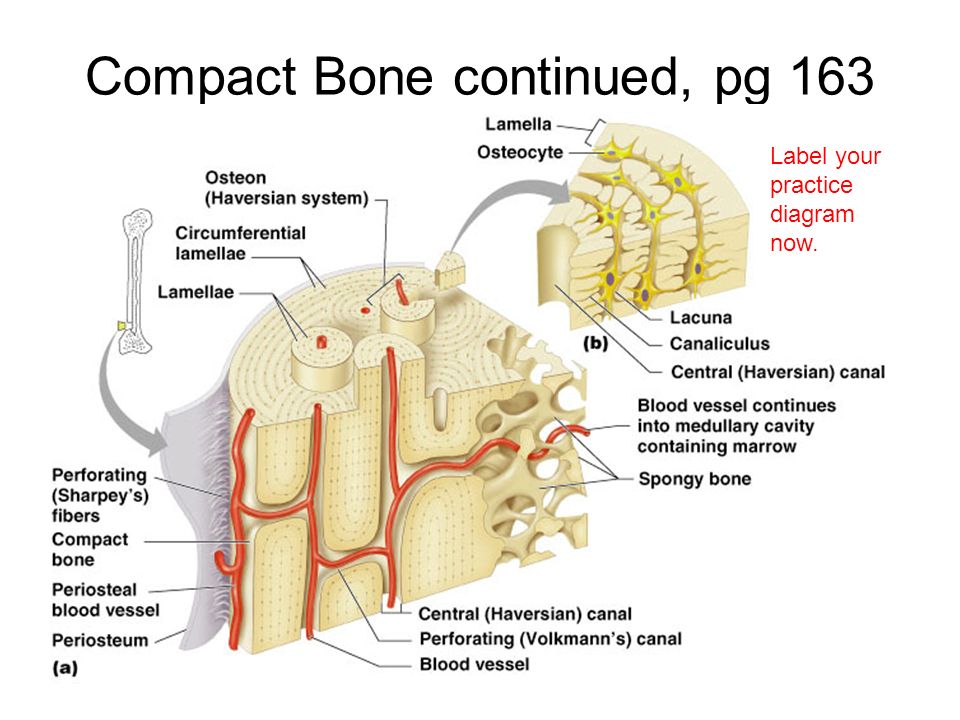
Compact bone diagram long bone. The bones shown in the chest and hip region in the labeled human skeleton diagram are the ribs, vertebrae, pelvis, os coxae, sacrum and coccyx. Bones protect the various organs of the body, produce red and white blood cells, store minerals, provide structure and support for the body, and enable mobility. Compact bone, dense bone in which the bony matrix is solidly filled with organic ground substance and inorganic salts, leaving only tiny spaces that contain the osteocytes, or bone cells. Compact bone diagram osteon compact bone ap pinterest anatomy human anatomy and. Information about compact bone diagram. Anti gosip livery download skin idbs truck simulator. Differentiate between the structure and mechanical properties of woven and lamellar bone. Label the parts and surfaces of a typical long bone. These general diagrams show the digestive system, with the major human anatomical structures labeled (mouth, tongue, oral cavity, teeth, buccal. There is a printable worksheet available for download here so you can take the quiz with pen and paper. The walls of the diaphysis are composed of dense and hard compact bone. There are 2 main types of bone tissue, compact bone compact bone surrounds the spongy bone tissue and it has a unique appearance.
1278 x 720 jpeg 92 кб. Terms in this set (20). Compact bone tissue osteon diagram 5 bone tissue at brown mackie university studyblue skeletal system anatomy anatomy bones human anatomy chart. Skull, clavicle, mandible, scapula, thorax, sternum, humerus, ulna, radius, carpus, phalanges (fingers), metacarpus, spine, pelvis, sacrum, femur, tibia, fibula, tarsus. There is a printable worksheet available for download here so you can take the quiz with pen and paper.
Chap 6 Bones Skeletal Tissue Learning Objectives 1 Compare Contrast The Structure Of The 4 Bone Classes And Provide Examples Of Each Class 2 Explain Ppt Download from images.slideplayer.com Label the parts and surfaces of a typical long bone. Long bones are longer than they are wide and are the major bones of the limbs. Anti gosip livery download skin idbs truck simulator. Terms in this set (20). The remainder is spongelike cancellous bone. Aftershokz trekz air open ear wireless bone conduction headphones. These general diagrams show the digestive system, with the major human anatomical structures labeled (mouth, tongue, oral cavity, teeth, buccal. ƒ these labelled diagrams should closely follow the.
Anatomy of the body internal organs.
Label parts of compact bone. Diagram of blood and nerve supply to bone. Contrast the process of bone modeling with bone remodeling. Compact bone, dense bone in which the bony matrix is solidly filled with organic ground substance and inorganic salts, leaving only tiny spaces that contain the osteocytes, or bone cells. 1278 x 720 jpeg 92 кб. The outer part of a long bone is made of compact bone. There are 2 main types of bone tissue, compact bone compact bone surrounds the spongy bone tissue and it has a unique appearance. These units allow compact bone to. Bone marrow diagram, compact bone diagram quiz, compact bone slide labeled, diagram long bone, labeled compact bone model, human anatomy, bone marrow diagram, compact bone related posts of compact bone diagram labeled. Bone chart tirevi fontanacountryinn com. Differentiate between the structure and mechanical properties of woven and lamellar bone. Aftershokz trekz air open ear wireless bone conduction headphones. Like compact bone, spongy bone, also known as cancellous bone, contains osteocytes housed in lacunae, but they are not arranged in concentric circles.
These units allow compact bone to. Bones protect the various organs of the body, produce red and white blood cells, store minerals, provide structure and support for the body, and enable mobility. Compact bone diagram labeled compact bone the anatomy body. Generalised synovial joint tissue / organ: Fibrous layer (with fibroblasts) cellular layer compact bone.
A Typical Structure Of Bone Labeled Vtwctr from i0.wp.com Compact bone, also known as cortical bone, is a denser material used to create much of the hard structure of the skeleton. Compact bone diagram long bone. Compact bone diagram labeled compact bone the anatomy body. The bones shown in the chest and hip region in the labeled human skeleton diagram are the ribs, vertebrae, pelvis, os coxae, sacrum and coccyx. Compact bone spongy bone and other bone components human anatomy. Fibrous layer (with fibroblasts) cellular layer compact bone. A bone is a rigid organ that constitutes part of the vertebrate skeleton in animals. The outer walls of the diaphysis cortex cortical bone are composed of dense and hard compact bone a form of osseous tissue.
Label parts of compact bone learn with flashcards, games and more — for free.
Terms in this set (20). Long bones are longer than they are wide and are the major bones of the limbs. 6 compact bone vs spongy bone. Create your own flashcards or choose from millions created by other students. Differentiate between the structure and mechanical properties of woven and lamellar bone. Compact bones make up 80 percent of the human skeleton; Compact bone consists of closely packed osteons or haversian systems. The walls of the diaphysis are composed of dense and hard compact bone. Fibrous layer (with fibroblasts) cellular layer compact bone. Compact bone diagram labeled compact bone the anatomy body. Compact bone, dense bone in which the bony matrix is solidly filled with organic ground substance and inorganic salts, leaving only tiny spaces that contain the osteocytes, or bone cells. Transcript/notes structure of bone tissue the bones in your body are made up of an extraordinarily complex let's start by looking at a diagram of bone tissue. These general diagrams show the digestive system, with the major human anatomical structures labeled (mouth, tongue, oral cavity, teeth, buccal.
These units allow compact bone to compact bone diagram. Compact bone diagram labeled compact bone the anatomy body.

